Biomechanical Comparison of Different Angles of K-wire Fixation Configuration for Management of Proximal Phalanx Fracture by Syringe External Fixators
DOI:
https://doi.org/10.56929/jseaortho-2026-0275Keywords:
Proximal Phalanx fracture, K-wire fixation, Syringe external fixation, Biomechanical comparison studyAbstract
Purpose: To optimize the K-wire fixation configuration for managing proximal phalanx fractures using Syringe External Fixators.
Methods: In this biomechanical comparison study, 48 sawbone models of proximal phalanx fractures stabilized with syringe external fixators were tested across eight different K-wire configurations (Groups A–H). Configuration included parallel, nonparallel, or combined patterns at angles of 0°, 30°, or 45°. The models were underwent longitudinal compression and pull-out tensile tests. Data were analyzed using one-way analysis of variance (ANOVA) to overall group comparison and independent t-test for pairwise comparisons.
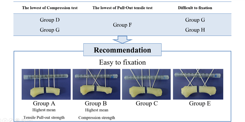
Results: Compression testing revealed that Group B (two parallel and two crossed K-wires) exhibited the highest mean ultimate strength (11.82 N). In contrast, Group D (two parallel and two crossed K-wires at varying angles) and G (four crossed K-wires) demonstrated the lowest strengths (5.49 N and 5.91 N, respectively). Although pairwise comparison between the highest- and lowest-strength groups showed a significant difference (p = 0.004), no statistically significant difference was observed across the eight groups in compression testing (p = 0.062). In pull-out testing, Group A (four parallel K-wires) displayed the highest mean ultimate strength (72.14 N), while group F (four cross-K-wires) showed the lowest (32.76 N). Pairwise comparison between these groups showed no statistically significant differences (p = 0.083). Similarly, no statistically significant difference in the pull-out tensile strength was observed among groups (p = 0.235).
Conclusions: In proximal phalanx fractures stabilized syringe external fixators, nonparallel and parallel K-wire fixation showed not significantly biomechanical different in compression and pull-out tensile testing.
Metrics
References
Azar FM, Canale ST, Beaty JH. Campbell's Operative Orthopaedics. 14th ed. Elsevier; 2020.
Yousaf O, Yousaf IS, Giladi AM, et al. Syringe External Fixator: An inexpensive static-to-dynamic treatment for comminuted intra-articular phalangeal fractures. Tech Hand Up Extrem Surg 2020;24:126-30. DOI: https://doi.org/10.1097/BTH.0000000000000280
Godwin Y, Arnstein PM. A cheap, disposable external fixator for comminuted phalangeal fractures. J Hand Surg Br 1998;23:84-5. DOI: https://doi.org/10.1016/S0266-7681(98)80227-1
Margić K. External fixation of closed metacarpal and phalangeal fractures of digits. A prospective study of one hundred consecutive patients. J Hand Surg Br 2006;31:30-40. DOI: https://doi.org/10.1016/J.JHSB.2005.09.013
Thomas RK, Gaheer RS, Ferdinand RD. A simple external fixator for complex finger fractures. Acta Orthop Belg 2008;74:109-13.
Dailiana Z, Agorastakis D, Varitimidis S, et al. Use of a mini-external fixator for the treatment of hand fractures. J Hand Surg Am 2009;34:630-6. DOI: https://doi.org/10.1016/j.jhsa.2008.12.017
Shah J, Rahman O, Ahmad R, et al. Phalangeal fractures- management by cost effective syringe fixators. JRMC [Internet]. 2015;19:133-5. Available from: https://www.journalrmc.com/index.php/JRMC/article/view/275.
Atthakomol P, Wangjiraphan N, Krudtong S, et al. Pull-out strength of 0°/30° Kirschner wire syringe external fixators with and without polymer augmentation: a biomechanical study. J Med Assoc Thai 2015:98:82-7.
Downloads
Published
How to Cite
Issue
Section
License
Copyright (c) 2026 The Royal College of Orthopaedic Surgeons of Thailand

This work is licensed under a Creative Commons Attribution-NonCommercial 4.0 International License.